

Marketing Campaign

0102030405
From Imaging to Therapy: How Radioactive Decay Shapes Clinical Use
2026-09-08
☢️ Not all radionuclides play the same role in nuclear medicine.
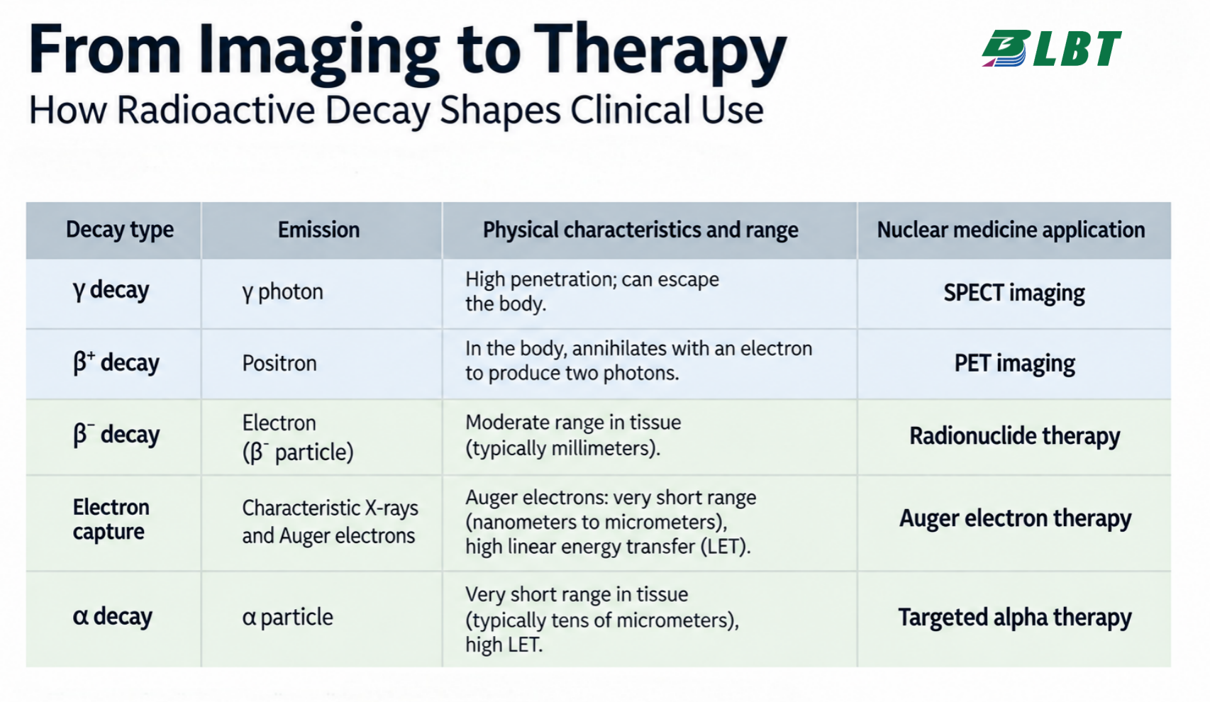
Whether a radionuclide is used for imaging or therapy depends largely on what it emits during radioactive decay—and how that radiation interacts with tissue.
A simple way to understand it:
Radiation that can escape the body can be detected for imaging.
Particles that deposit energy locally can be used for therapy.
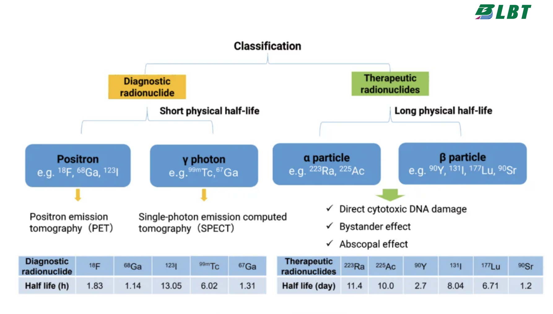
🔍 γ rays — SPECT imaging Gamma photons can travel out of the body and be detected externally, making radionuclides such as ⁹⁹ᵐTc and ¹²³I suitable for SPECT imaging.
📡 β⁺ decay — PET imaging Positron emitters such as ¹⁸F, ⁶⁸Ga, ¹¹C and ¹³N produce positrons that eventually annihilate with electrons, generating photon pairs detected by PET scanners.
Together, these radionuclides allow us to visualize physiological and molecular processes inside the body.
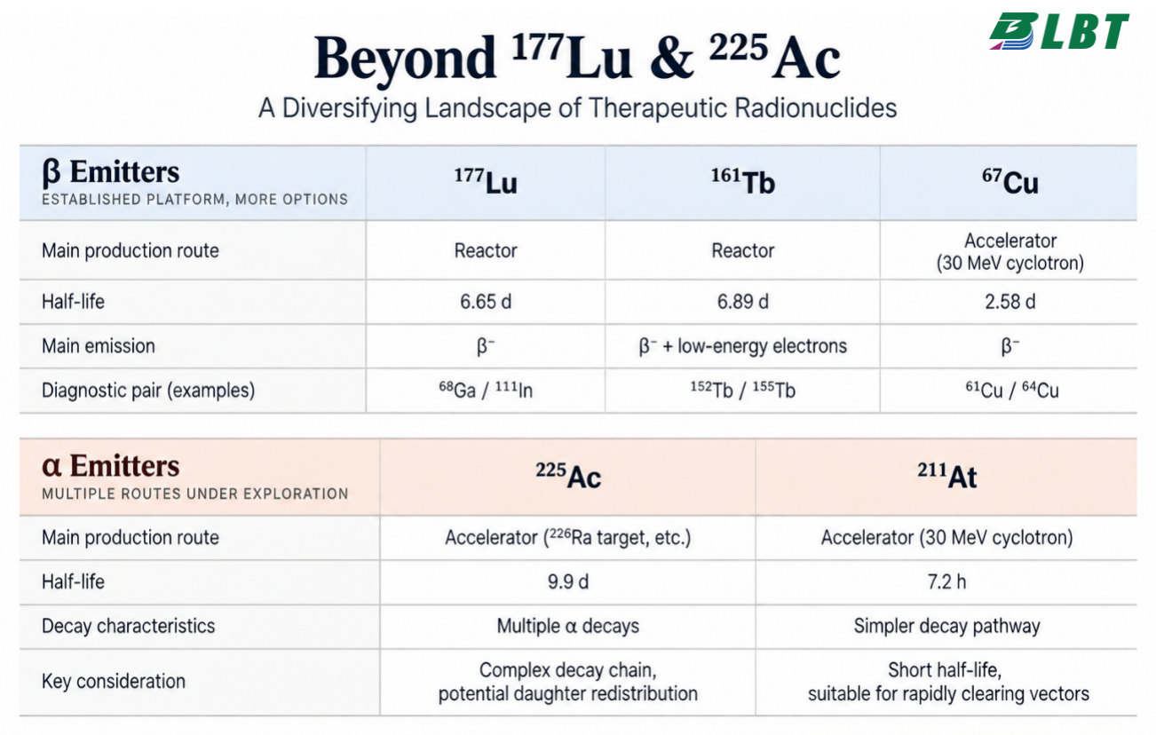
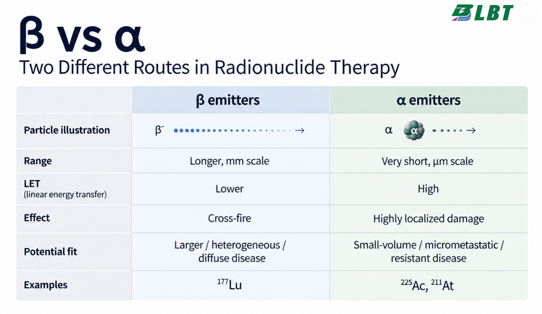
🎯 β⁻ particles — therapeutic radiation Beta particles travel over a relatively longer range in tissue, allowing radiation to affect both targeted and nearby cells—the cross-fire effect. ¹⁷⁷Lu and ⁹⁰Y are representative β⁻ therapeutic radionuclides.
⚡ α particles — short range, high energy deposition Alpha particles travel only a very short distance in tissue but deposit energy densely along their path. Radionuclides such as ²²³Ra, ²²⁵Ac and ²¹¹At are used or being investigated for targeted alpha therapy.
🔄 Different emissions. Different clinical roles.
γ / β⁺ → Imaging 🔍 β⁻ / α → Therapy 🎯
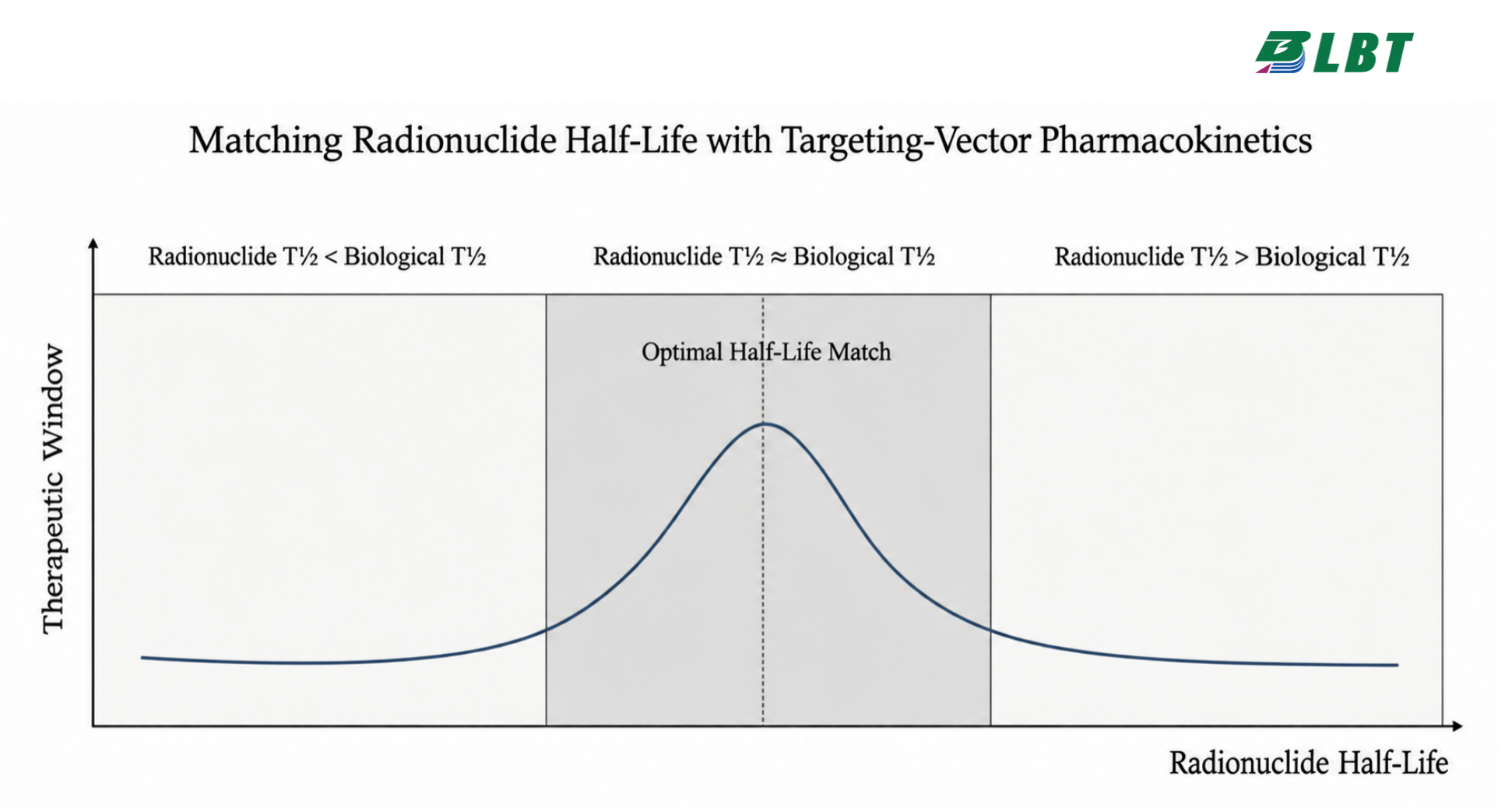
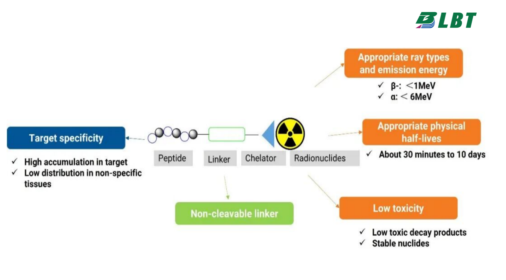
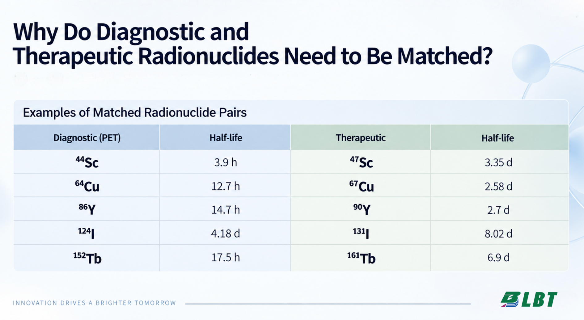
But even radionuclides within the same category are not interchangeable. Their range, energy, half-life and decay characteristics all influence how they can be used.
Ultimately, radionuclide selection starts with one question:
What do we want the radiation to do inside the body?